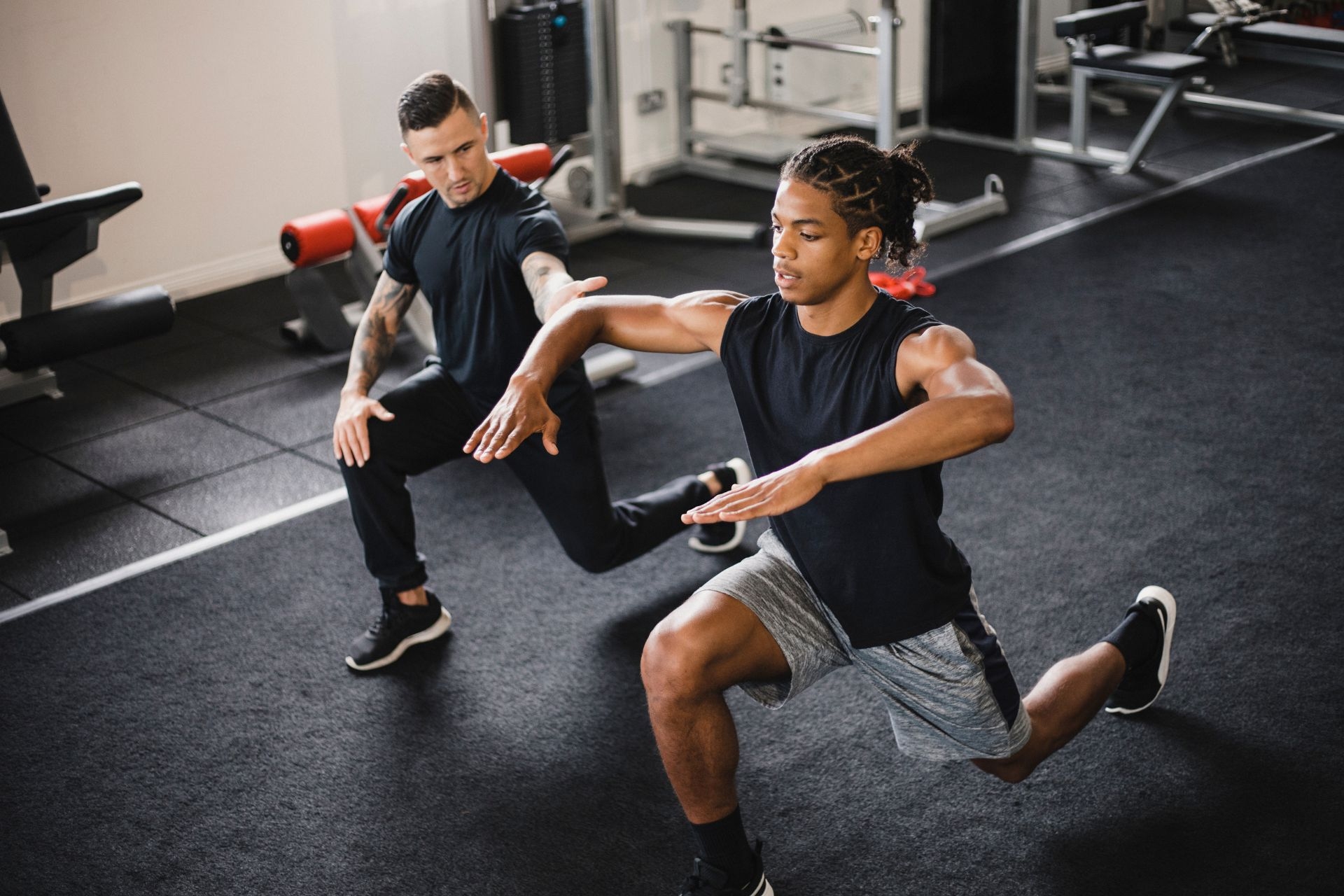
Muscle spindles play a crucial role in proprioception by detecting changes in muscle length and relaying this information to the central nervous system. These specialized sensory receptors are located within the muscle belly and are sensitive to stretch, allowing them to provide feedback on the position and movement of the body in space.
Gamma motor neurons are responsible for regulating muscle spindle sensitivity. By adjusting the tension in the intrafusal muscle fibers within the spindle, gamma motor neurons can modulate the responsiveness of the spindle to changes in muscle length. This mechanism helps to fine-tune proprioceptive feedback and ensure accurate perception of body position.
Pediatric PT Approved Early Riding Toys! The post Our Favorite Early Riding Toys appeared first on DINOSAUR PHYSICAL THERAPY.
Posted by on 2024-03-01
Alternative Seating Options for Children! The post Alternative Seating Options appeared first on DINOSAUR PHYSICAL THERAPY.
Posted by on 2024-02-12
Pediatric PT Approved Cold Weather Shoe Recommendations! The post Shoe Recommendations for Children: Cold Weather Edition appeared first on DINOSAUR PHYSICAL THERAPY.
Posted by on 2024-01-31
Stair Climbing Tips and Tricks! The post Stair Climbing Tips and Tricks appeared first on DINOSAUR PHYSICAL THERAPY.
Posted by on 2021-06-03
Muscle spindles detect changes in muscle length through the stretching of their sensory nerve endings, known as primary afferent fibers. When a muscle is stretched, the spindles are activated, sending signals to the spinal cord and brain about the extent of the stretch. This information is crucial for coordinating movement and maintaining posture.
The stretch reflex is a rapid, involuntary response to muscle stretch that is mediated by muscle spindles. When a muscle is stretched, the spindles detect the change in length and send signals to the spinal cord, which in turn triggers a reflex contraction of the muscle to resist further stretching. This reflex helps to protect the muscle from overstretching and plays a key role in maintaining stability and balance.
Muscle spindles differ from Golgi tendon organs in terms of their function. While muscle spindles detect changes in muscle length and contribute to proprioception, Golgi tendon organs are sensitive to changes in muscle tension and provide feedback on muscle force. Both types of sensory receptors work together to regulate muscle activity and prevent injury.
Muscle spindles have the ability to adapt to changes in muscle length over time through a process known as sensory adaptation. This allows the spindles to remain responsive to dynamic changes in muscle length while filtering out constant or repetitive stimuli. By adapting to different movement patterns and loads, muscle spindles help to maintain accurate proprioceptive feedback.
Muscle spindles contribute to motor control and coordination by providing real-time feedback on muscle length and movement. This information is essential for adjusting muscle activity and coordinating the contraction and relaxation of muscles during various tasks. By integrating sensory input from muscle spindles with other sensory information, the central nervous system can effectively regulate movement patterns and ensure smooth and coordinated motor control.
Palpation can be utilized as a valuable tool in assessing the diaphragm for breathing dysfunction by applying gentle pressure to the abdominal area to feel for any abnormalities or restrictions in movement. By palpating the diaphragm, healthcare providers can detect any signs of decreased mobility, tenderness, or asymmetry that may indicate dysfunction. Additionally, palpation can help identify trigger points or areas of increased tension within the diaphragm muscle, which could be contributing to breathing difficulties. By incorporating palpation into a comprehensive assessment of the diaphragm, healthcare providers can gather valuable information to guide treatment strategies and improve respiratory function. This hands-on approach allows for a more thorough evaluation of the diaphragm's function and can help identify underlying issues that may be impacting breathing patterns.
When palpating the rhomboid muscles for tension or spasm, there are several methods that can be utilized. The therapist can start by gently pressing their fingers along the medial border of the scapula, feeling for any areas of tightness or knots. They can also use a cross-fiber friction technique to help release any adhesions or trigger points within the muscle. Additionally, applying deep pressure along the muscle fibers while the client performs active movements, such as shoulder retractions, can help identify areas of increased tension or spasm. By combining these various palpation techniques, the therapist can effectively assess the condition of the rhomboid muscles and develop an appropriate treatment plan to address any issues present.
Palpation can be a valuable tool in identifying a hamstring strain by allowing the healthcare provider to feel for specific signs of injury such as tenderness, swelling, and muscle tightness. By applying pressure to the affected area, the provider can assess the extent of the strain and determine the location of the injury. Additionally, palpation can help differentiate between a hamstring strain and other possible causes of pain in the same region, such as a muscle tear or tendonitis. The provider may also use palpation to assess the overall condition of the surrounding muscles and tissues to develop an appropriate treatment plan for the patient. Overall, palpation plays a crucial role in the diagnostic process of hamstring strains by providing valuable information about the nature and severity of the injury.
To palpate the sacroiliac joint for dysfunction, a healthcare provider can have the patient lie on their side with the affected side facing up. The provider can then locate the posterior superior iliac spine and move their fingers inferiorly towards the sacrum until they reach the sacroiliac joint. By applying gentle pressure and assessing for tenderness, asymmetry, or restricted motion, the provider can determine if there is dysfunction present in the joint. Additionally, the provider may perform specific tests such as the Gaenslen's test or the thigh thrust test to further evaluate the sacroiliac joint for dysfunction. It is important for the provider to have a thorough understanding of the anatomy and biomechanics of the sacroiliac joint to accurately assess for dysfunction and develop an appropriate treatment plan.
Palpation is a valuable technique used by healthcare professionals to assess the integrity of the Achilles tendon. By applying pressure and feeling along the length of the tendon, practitioners can detect any abnormalities such as swelling, tenderness, or nodules that may indicate a potential injury or condition. Palpation can also help determine the extent of damage, such as the presence of a partial or complete tear, as well as assess the overall health and flexibility of the tendon. Additionally, comparing the Achilles tendon on both sides can provide valuable information about symmetry and potential imbalances. Overall, palpation plays a crucial role in the evaluation of the Achilles tendon and can guide further diagnostic and treatment decisions.
Palpation can be utilized to assess pelvic alignment for signs of asymmetry by carefully feeling for any irregularities, discrepancies, or imbalances in the bony structures, muscles, and soft tissues surrounding the pelvis. By applying gentle pressure and using specific hand placements, a skilled practitioner can detect differences in the position, movement, and tension of the pelvic bones, sacroiliac joints, hip joints, and surrounding ligaments. Additionally, palpation can help identify any areas of tenderness, inflammation, or restricted mobility that may indicate pelvic misalignment. By systematically evaluating the pelvis through palpation, healthcare providers can gather valuable information to guide treatment strategies and address any underlying issues contributing to asymmetry.