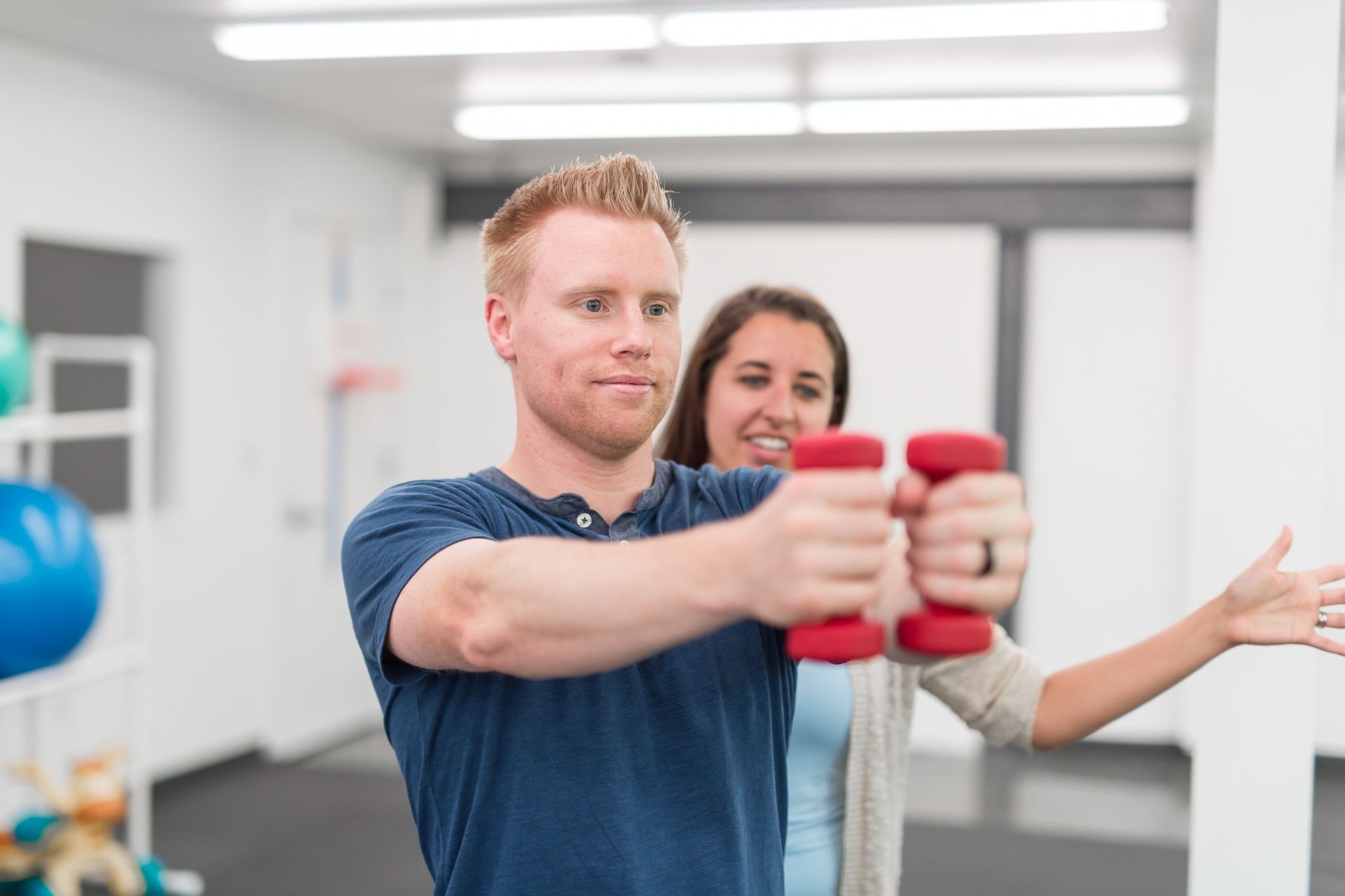
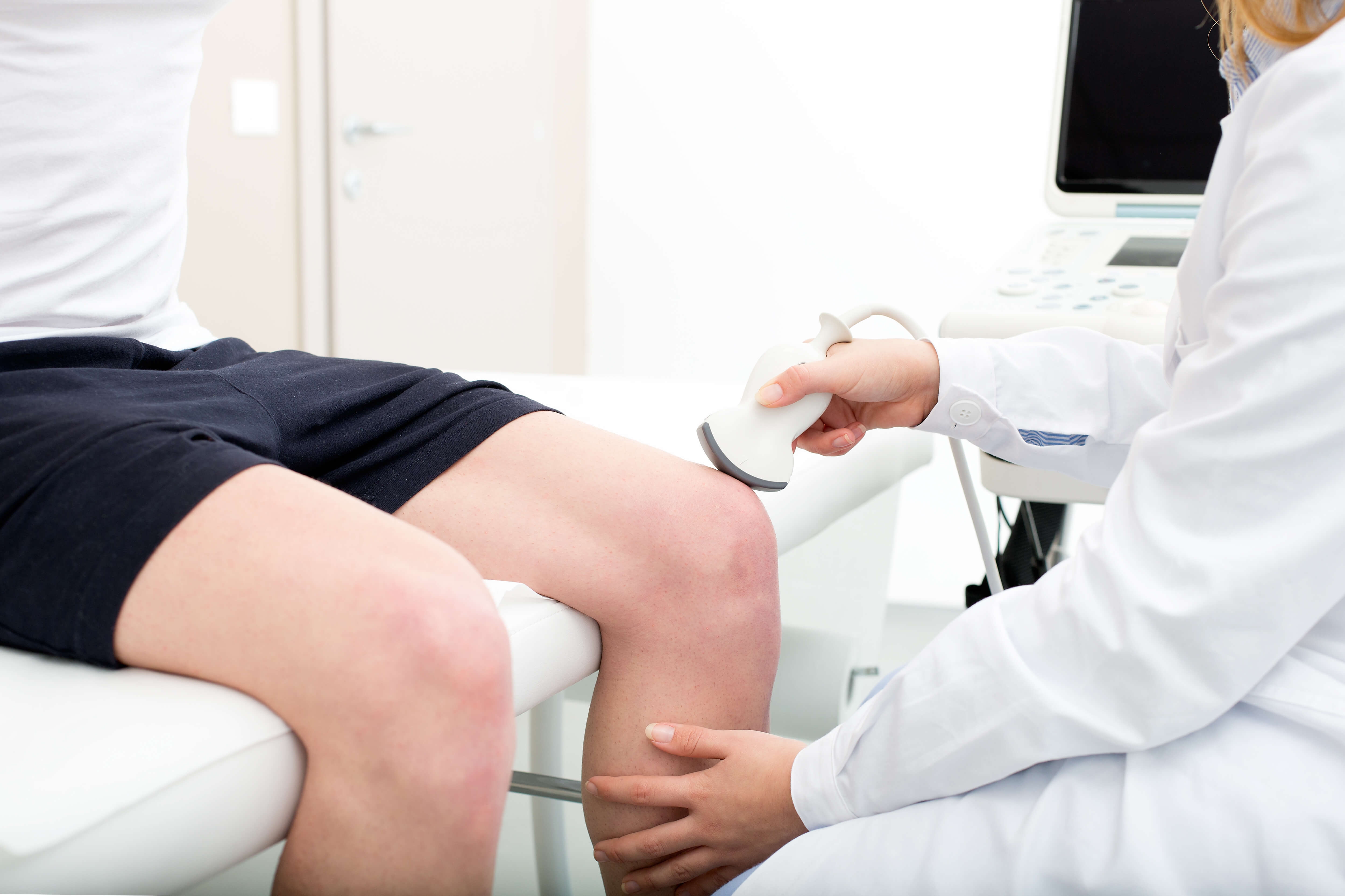
Active Release Techniques
How does Active Release Techniques help with repetitive strain injuries?
Active Release Techniques (ART) is highly effective in treating repetitive strain injuries by targeting the specific muscles, tendons, ligaments, and nerves affected by the repetitive movements. ART involves a combination of precisely directed tension and specific movements to break up adhesions and scar tissue, allowing for improved blood flow and range of motion in the affected area. By addressing the root cause of the injury, ART can help alleviate pain and restore function in individuals suffering from repetitive strain injuries.