September 7, 2024
Ultrasound Therapy: What Is It, Just How It Functions, Advantages
Ultrasound Therapy For Persistent Reduced Pain In The Back Diagnostic ultrasound is usually defined by the facility regularity of the pulses (commonly in the 2-- 12 MHz array), which is typically a regularity integral to the thickness of the ceramic crystal. As the pressure amplitude, the frequency, or the propagation size is enhanced, the ultrasound wave can misshape, which might ultimately lead to a suspension or shock in the waveform. In regard to bioeffects, boosting regularity, nonlinear acoustic distortion, or pulse length can boost home heating and improve some nonthermal mechanisms, e.g., radiation pressure. Decreasing frequency enhances the probability of cavitation and gas body activation. Raising power or intensity has a tendency to enhance the possibility and magnitude of all bioeffects mechanisms. Restorative ultrasound tools may make use of brief ruptureds or continuous waves to provide efficient ultrasonic power to cells.- Although ultrasound treatment is usually safe, the stick should not be kept in one area for too lengthy at particular regularities.
- To guarantee your safety, both of our ultrasound treatment tools are outfitted with a system for acknowledging correct call in between the ultrasound head and the skin.
- An efficient version-- though less current than Powersonic-- is Mio-Sonic.
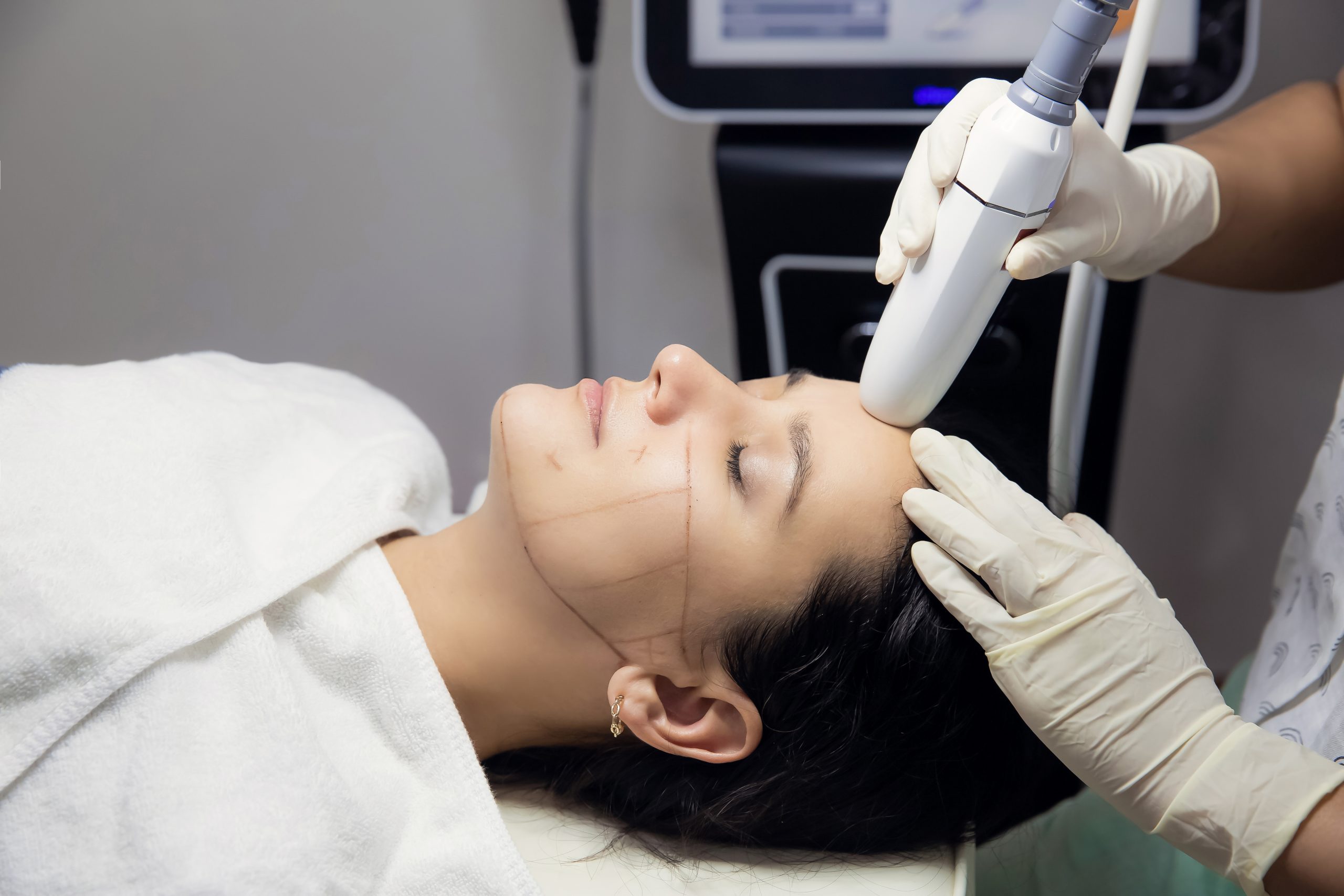
- The probe is normally a handheld device with piezoelectric crystals that produce the ultrasound waves.
How deep does ultrasound treatment permeate?
Restorative ultrasound in physical therapy is rotating compression and rarefaction of sound waves with a frequency of 0.7 to 3.3 MHz. Maximum power absorption in soft cells occurs from 2 to 5 centimeters. Intensity lowers as the waves permeate much deeper.
What Are The Dangers Of Therapeutic Ultrasound?
This is one evidence-based formula that a lot of professionals make use of. It sends or shoots out an ultrasound wave from the soundhead. You can have a continual wave where it's 100% moving at all times, and after that if you look listed below on the representation, you can have what is called a pulse wave or a pulsed wave. A pulsed-wave provides you a hidden period or a pause in between ultrasound waves. Depending on what your setting is for your ultrasound, you can utilize a continuous wave or you can make use of a pulsed wave to promote different physical processes in the body.Can Ultrasound Harm?
When methodologically flawed trials were left out, there were few RCTs that checked out ultrasound and those RCTs offered little clinical proof for the effectiveness of healing ultrasound. The application of the exclusion criteria and methodological filters caused the removal of all except 10 professional ultrasound trials from today testimonial. 8 studies showed that active ultrasound is no more useful than sugar pill ultrasound for the treatment of people with discomfort or soft cells injury. Couple of generalizations can be drawn from the 2 tests in which active ultrasound was discovered to be above placebo ultrasound, provided their heterogeneity and omission of essential details.Just How Does Restorative Ultr
Before starting the treatment, the technician will evaluate the skin for any kind of infections, burns, or active wounds. If your skin is clear, after that the service technician will apply a hypoallergenic gel or lotion to your skin. This will assist keep air from coming between your skin and the stick and likewise aid perform the ultrasound waves to the tissues. The ultrasound will certainly then be used on your skin by the specialist for regarding 10 mins in a rubbing activity. Ultrasound-- or ultrasonography-- is an imaging strategy made use of not just during pregnancy https://nyc3.digitaloceanspaces.com/health-nutrition/healthy-habits/wrinkle-reduction/nonsurgical-facelift-what-it-is95395.html however also for lots of clinical procedures. Ultrasound physical therapy is a branch of ultrasound, together with analysis ultrasound and pregnancy imaging. A radiologist can determine the size and color versus other close-by body organs to compare them. Projections or areas externally could show cysts or strong masses. A radiologist may additionally try to find enlarged (dilated) blood vessels or bile ducts. Your sonographer will send out the images from your test to a radiologist.Social Links